What is Symblepharon?
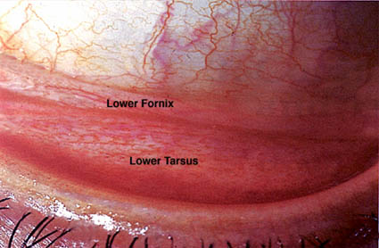
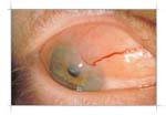
A symblepharon is a fibrous tract that connects bulbar conjunctiva toconjunctiva on the eyelid.
 |
![Symbol of [describe the symbol, e.g., a company logo or a specific graphic, and any embedded text in the image]](/topics/190-Overview/Image/symble1.jpg) |
| Symblepharon, Normal Fornix |
Symblepharon, Upper Fornix |
When do symblepharon occur?
- Cicatricial pemphigoid; auto-immune disease which affects mucus membranes such as the mouth and oral pharynx, conjunctiva, nares and genitalia
- Atopic keratoconjunctivitis: This is a hypersensitivity to environmental allergies including asthma, rhinitis, dermatitis and eczema.
- Trichiasis: A lid abnormality in which eyelashes are misdirected towards the eyeball. These misdirected lashes could often be the lead to of scarring.
- Toxic epidermal necrosis (TEN): A potentially life-threatening disorder which is commonly drug-induced.
- Stevens Johnson Syndrome
- Characteristics:
- Equal age and sex distribution
- disease associated with a 5-15% mortality rate
- Ocular involvement in 50%
- Associated with various infections and medications most notably sulfa
Ocular involvement in 50%
- Pathogenesis:
- Angiitis-->Erythematous lesion-->Bullae-->Rupture-->Scar
- Prodrome:
- Fever, chills, and headache
- 7 days later bullous mucosal lesions develop
- Sequelae:
- Problems are related to the destruction of Goblet cells and a lack of conjunctival mucus which leads to keratinization and scarring.
- Lid scarring with symblepharon
- Corneal scarring
- Keratitis sicc
- Burns
- Erythema multiforme: This is an acute multi-cutaneous hypersensitivity reaction
What are the treatment options?
- Why use Amniotic membrane? Because it:
- facilitates epithelialization
- maintains normal epithelial phenotype (with goblet cells when performed on conjunctiva ),
- reduce inflammation, vascularization and scarring.
The use of {human} amniotic membrane for the surgical treatment of an ocular surface disorder was originally reported by de Rotth 16 in 1940. During the 1990s, the role of amniotic membrane transplantation in treating a variety of ocular surface defects and abnormalities has been revived.
Chemical Burns
|
Pterygium |
 |
 |
Corneal/Sclera Ulceration &
Peforations |
Tumors |
 |
 |
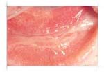
| Stevens-Johnson's Syndrome |
|


![Symbol of [describe the symbol, e.g., a company logo or a specific graphic, and any embedded text in the image]](/topics/190-Overview/Image/symble1.jpg)


